


Welcome to Wide-field Kerr microscopy and magnetometry
The evico magnetics GmbH was founded in 2006 as spin-off of the Leibniz Institut for Solid State and Materials Research (IFW) Dresden.
The main products are: (i) Advanced magneto-optical wide-field Kerr microscope systems for the visualization of magnetic domains and magnetization processes in all kinds of magnetic materials. The Kerr microscopes also serve as magneto-optical magnetometers for the sensitive and local measurement of hysteresis loops by MOKE magnetometry. (ii) High Pressure Milling Vials with a gas temperature monitoring system for the synthesis of magnetic powders and hydrogen storage materials.
Highlights and Applications
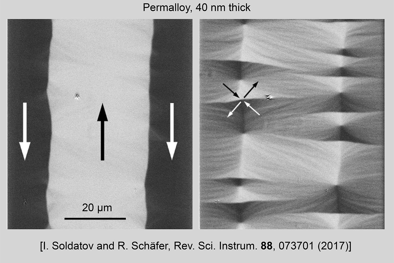
Multicomponent imaging in real time
Separated or combined imaging of longitudinal, transverse and polar magnetization components by sophisticated LED light source, computer-controlled and in real time
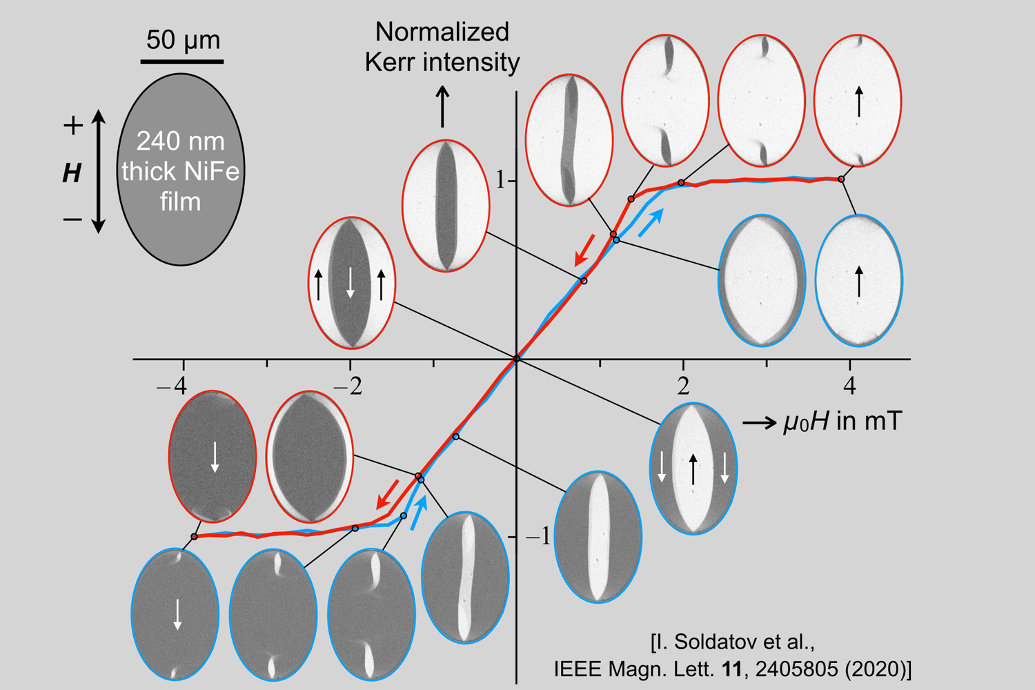
MOKE magnetometry
Our Kerr microscope serves as a fast and sensitive MOKE magnetometer. The domains responsible for the magnetization curve are supplied in real time
High-resolution Kerr microscopy
A lateral resolution of ~180 nm can be achieved
Quantitative Kerr microscopy
Magnetization vector fields of soft magnetic specimens and their dynamics can be quantitatively measured
Sample stabilization
Sample drift in all spatial directions is in-situ and at real time compensated to guarantee perfect domain images
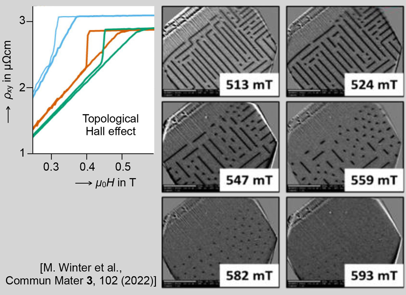
Combination with transport measurement
Domain imaging and magnetometry can be in-situ combined with real-time electrical transport measurements
Current-induced domain generation and motion in PMA films with DMI
The first experimental evidence for domain wall motion in the racetrack memory and skyrmionic-bubble generation by electric currents was provided in evico magnetics Kerr microscopes
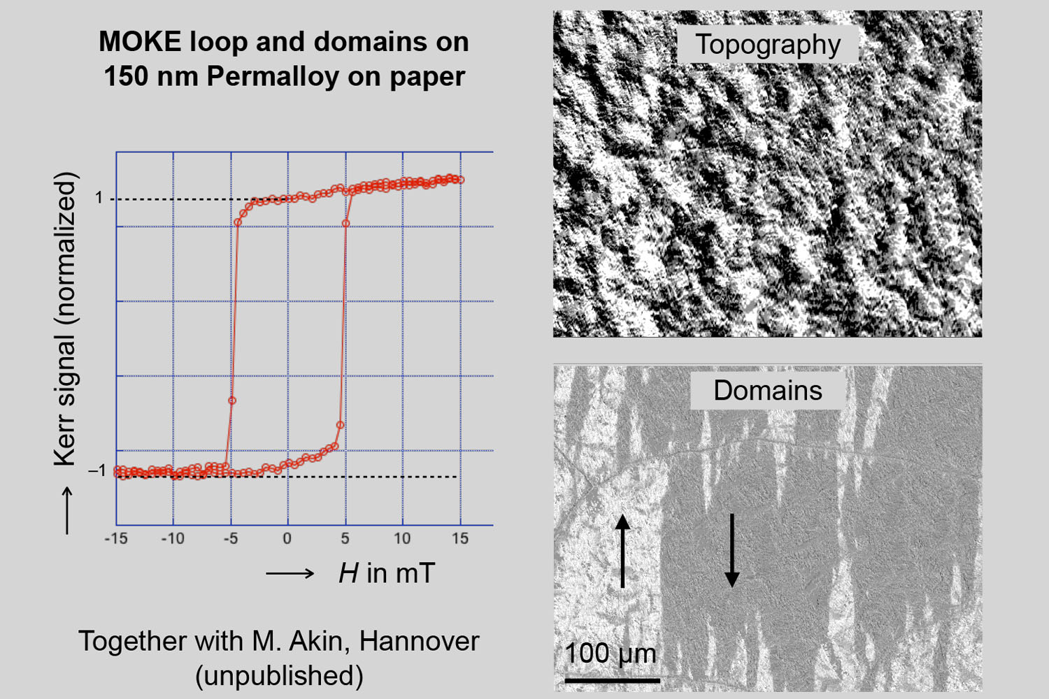
Ultimate in-plane sensitivity
A unique LED lamp illumination scheme makes it possible to enhance the Kerr sensitivity of in-plane magnetized materials, allowing for domain contrast and magnetometry on specimens with extremely rough surface
Time-resolved domain imaging
Tools for single-shot imaging up to power frequencies or stroboscopic imaging at higher frequencies are offered
Overview Kerr microscopy
First get an overview of the domain pattern (30 mm image size) before zooming into the pattern at high resolution – combined overview / high-resolution Kerr microscopy offers clear advantages in domain imaging and MOKE magnetometry
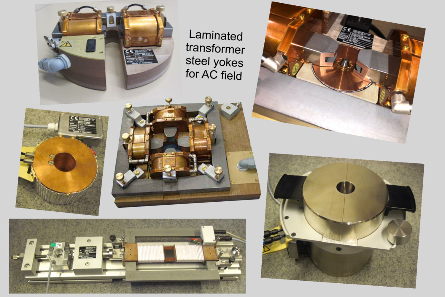
Magnetic field options
Numerous electromagnets are offered, allowing rotatable in-plane, perpendicular, or superimposed magnet fields beyond the Tesla range. Ribbon and sheet specimens can be imaged in a single-sheet magnet in combination with inductive (fluxmeter-based) dynamic hysteresis measurements
Polar real-time imaging
Domains and magnetization processes in pma media are easily accessible at high resolution, isolated objects down to the 50 nm size range can still be seen
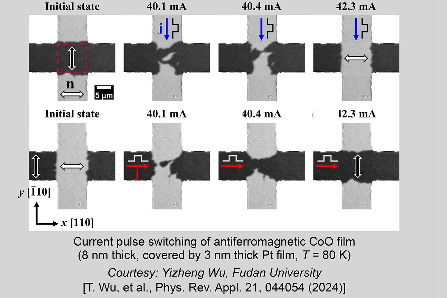
Domain imaging of antiferromagnetic films
Applying the Linear Magnetic Birefringence effect in an evico magnetics Kerr microscope allows to image domains in antiferromagnetic materials
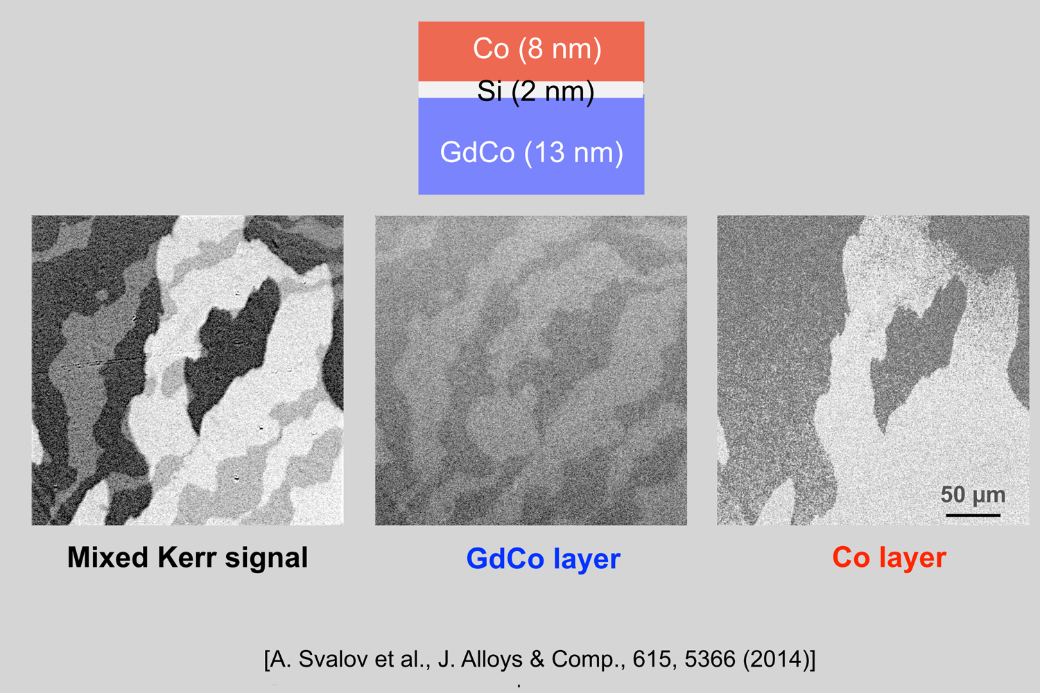
Depth sensitive domain imaging
Custom-designed imaging tools, like layer selective Kerr microscopy for the examination of magnetic multilayer specimens, are provided
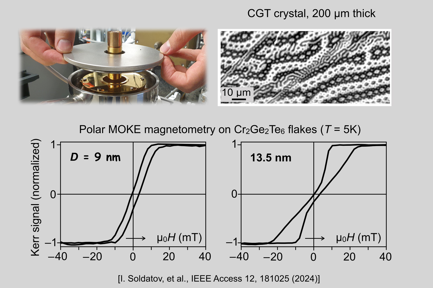
Low-and high temperature microscopy and magnetometry
Domain imaging and magnetometry can be performed in a temperature range between 4 K and 850 K in optical cryostat and heating stages, compatible with in-plane and out-of-plane magnets
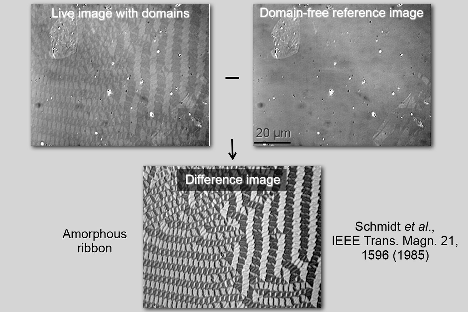
Image processing
Background subtraction eliminates topographic contrast, leaving just magnetic domain contrast that can be enhanced digitally
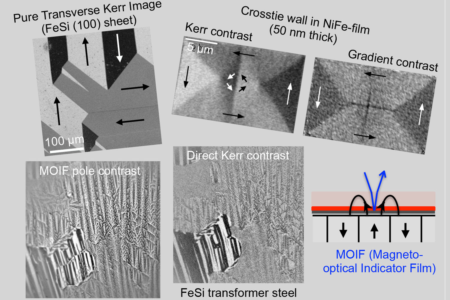
Sophisticated magneto-optical microscopy
evico magnetics microscopes are more than Kerr microscopes: Magnetic poles can be visualised by MOIF Microscopy, magnetization gradients show up in the Schaefer-Hubert Effect, intensity-based effects offer alternatives to conventional imaging – ask us
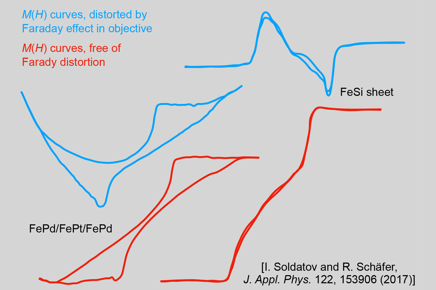
Faraday-free magnetometry and microscopy
MOKE magnetometry and microscopy may suffer from Faraday rotations in the objective lens, which are caused by the applied magnetic field or by stray fields emerging from the sample edges and which are superimposed to the Kerr signal. We offer solutions to avoid that – ask us
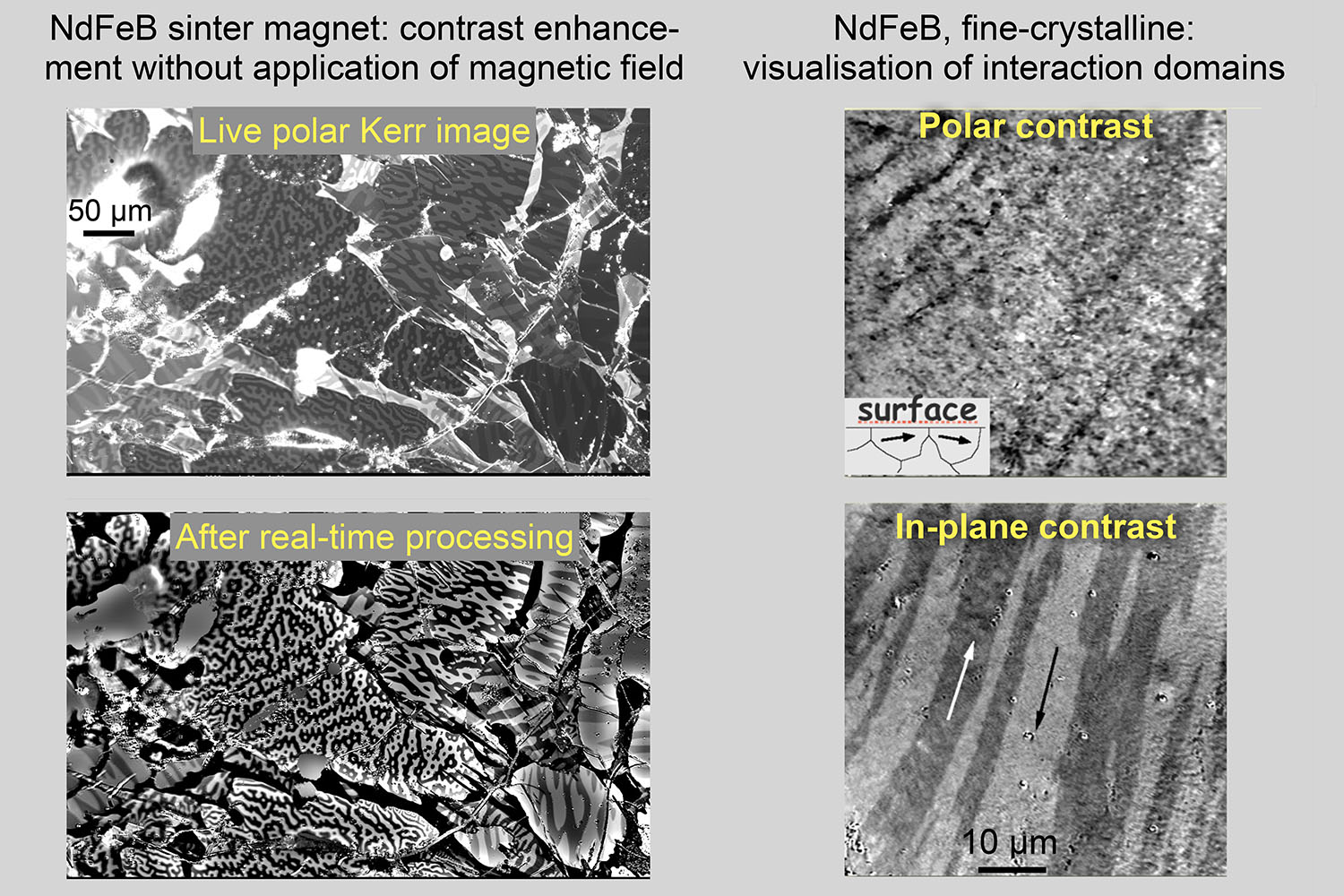
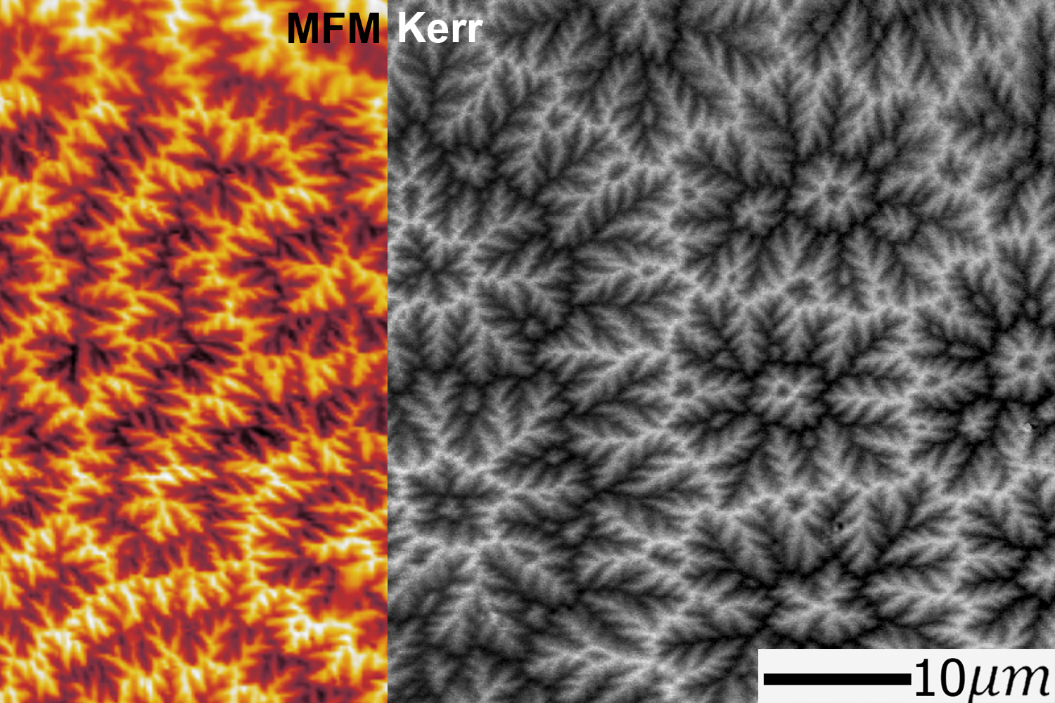
Advanced imaging of permanent magnet materials
Left: High-quality domain images can be obtained in quasi real-time without the need to apply saturation fields for background subtraction.
Right: surprisingly, wide in-plane magneto-static interaction domains appear in fine-crystalline material after contrast separation. The upper image shows the same domain state in a regular polar Kerr micrograph
Advanced imaging of permanent magnet materials
Wide in-plane magneto-static interaction domains appear in fine-crystalline material upon field application