

Kerr microscopy is based on the magneto-optical Kerr effect, which causes a rotation of linearly polarized light when reflected by a magnetic material.
.
The Kerr effect can be understood classically on the basis of the Lorentz concept: The electric field vector E of the incident linearly polarized light causes an oscillation of the elections in the material along the plane of incidence. Due to the magnetization vector m, the oscillating electrons experience a Lorentz force (m x E), which causes the plane of oscillation to rotate. The electric field vector of the reflected light emitted by the oscillating electrons is therefore composed of a normal reflected component (AN ) and a transverse Kerr component (AK ), which leads to the aforementioned Kerr rotation. The rotation can be clockwise or
anti-clockwise, depending on the direction of magnetization.
.
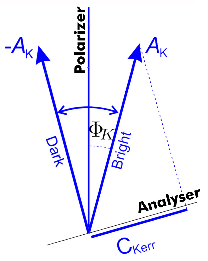
If the light emitted by the domain phases with different rotation angles is blocked differently using an analyser, a domain contrast is produced in an optical polarization microscope. If there is a phase shift between the normal and Kerr amplitude, the ellipticity can be compensated for by a phase shifter (compensator, e.g. quarter-wave plate) in front of the analyser